Why bone density matters in your care
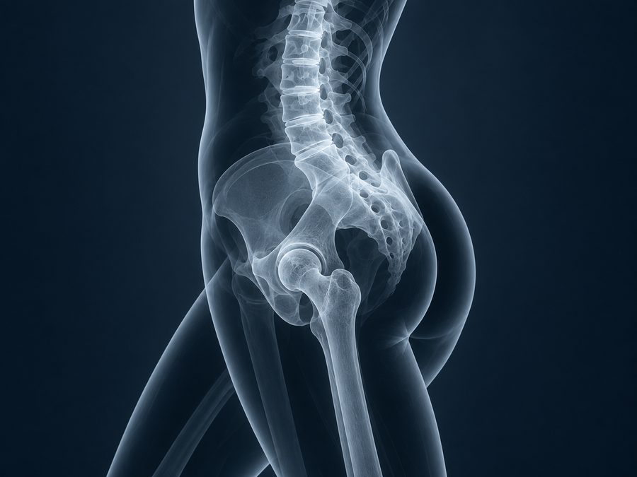
Bone loss can progress without obvious symptoms. In HLC care, the result helps connect bone health with strength, balance, nutrition, hormonal history and medication use. It can change the priority of resistance exercise, fall prevention or a specialist assessment.
The decision to scan depends on your risk profile. A previous low-trauma fracture, menopause, long-term use of medicines that affect bone, or a relevant medical condition may make the question more important.
How the examination works
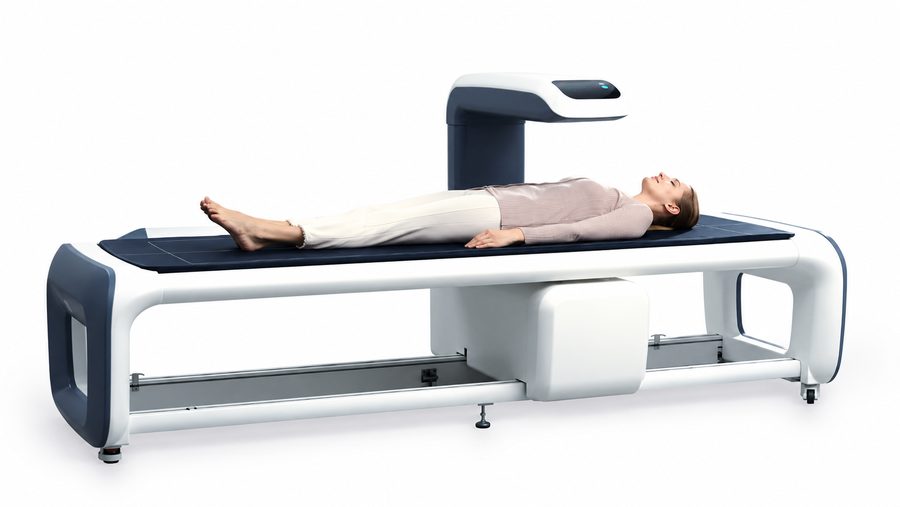
The team reviews your history and positions you for each region. A support under the legs may be used for the spine, while the foot is positioned differently for the hip. Keeping still helps the measurements remain reliable. DXA is an open examination rather than a tunnel scan.
A scan commonly takes around 10–30 minutes. This is general examination guidance; the clinic confirms your appointment length, measured sites and whether the result consultation is part of the booking.
What the numbers mean
Bone mineral density is the measured amount of mineral projected over an area of bone. A T-score compares it with a young-adult reference; a Z-score compares it with an age-appropriate reference. Which score guides interpretation depends on age and clinical context.
In postmenopausal women and men aged 50 or older, T-scores are generally used for bone-density classification. In younger adults, Z-scores usually provide the more appropriate comparison. The score does not, by itself, tell us whether or when a fracture will happen.
Before your visit
Bring previous bone-density reports and a list of medicines and supplements. Tell the team about possible pregnancy, recent contrast or barium examinations, fractures, implants or spinal surgery. Wear comfortable clothing without metal near the scan area and follow the imaging centre’s instructions about calcium supplements.
Turning the report into next steps
Your clinician combines the result with fracture history and other risks, then discusses practical bone-health measures and whether further tests or treatment are needed. Repeat scans are most useful when they can influence care. Small changes between scans may reflect measurement variation, so comparable equipment and the facility’s precision matter.